Electron Microscope Applications and Preparation of Samples
Due to their ability to achieve resolution of 50 pm and magnify the specimen for up to 10 million times, electron microscopes are of vital importance for many fields of science and research, especially biology, medicine and life sciences. However, they are also an indispensable piece of equipment in nanometrology, classification of materials, device testing, etc. but they are also used in industry for a number of reasons including quality control.
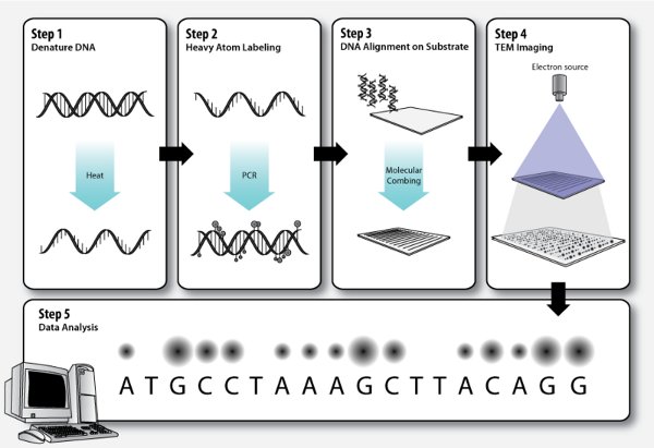
In order to make the most of electron microscope, the samples that are observed under the electron beam must be properly prepared. The preparation of samples depends on the type of electron microscope used, specimen and analysis. It may involve one or multiple of the following preparation techniques:
Conductive coating. The method refers to extremely thin coating of a material that is electrically conducting such as gold, platinum, palladium or chromium with an aim to prevent static electric fields from accumulating at the specimen during imaging.
Cyrofixation. It involves rapid freezing of the specimen to extremely low temperatures to create a snapshot of the specimen’s solution. This allows imaging of biological specimens close to their native state.
Dehydration. The technique is used to remove water from the sample. Depending on the type of the microscope used, the removal of water with the use of acetone or ethanol can be followed by supercritical drying or infiltration with resin.
Embedding. It involves infiltration of the specimen with a resin in order to allow sectioning of the sample.
Freeze-fracture, freeze-etch. Samples prepared for imaging according to this technique are frozen rapidly (cyrofixed) and then fractured. The fractured sample is then typically “etched” by increasing the temperature to about -95 degrees Celsius. But while such sample is ready to be observed with the SEM, further processing is required to be able to observe it with the TEM.
Fixation. It involves preservation of a specimen with the use of various chemicals in order to prevent decay or deterioration, allowing the researcher to observe the specimen as if it would be in a living state.
Sectioning. Depending on the analysis required, the specimen may be sectioned in thin slices either with a diamond or glass knife.
Staining. In order to give particular structures of the sample a higher contrast, the sample may be stained with heavy metals. Staining can be performed before or after embedding and sectioning.
Preparation of the samples is an important part of electron microscopy. Unfortunately, it is often a time consuming and complicated procedure but it is inevitable for the specimens to withstand the conditions within an electron microscope. Samples are always viewed in a vacuum although the so-called environmental scanning electron microscope (ESEM) allows the use of wet samples. The ESEM applications, however, are currently still limited.