History of Electron Microscope

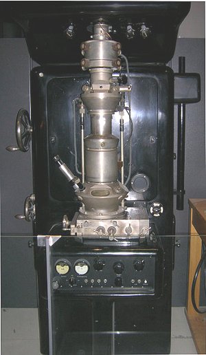
The first prototype of microscope which uses a beam of electrons rather protons (light) was constructed by the German scientists Max Knoll and Ernst Ruska in 1931. However, the world’s first electron microscope is sometimes considered the model that was built by Ruska in 1933 with a capacity to resolve to 50nm. However, both the microscope that Ruska built together with Knoll and the version that was later built by himself were primitive and unsuitable for practical applications.
Although Ruska (and Knoll) are credited with the invention of electron microscope, the patent was actually obtained by Reinhold Rudenberg, scientific director of Siemens in 1931. He became interested in alternative to optical microscope in 1930 when his son was diagnosed with poliomyelitis, a potentially life-threatening condition which was known to be caused by a viral infection. However, the virus could not be seen with the light microscope. Rudenberg was determined to develop a device which will make the tiny virus visible. As a former student of Emil Wiechert who played an important role in the discovery of the electron and as a friend of Hans Busch who was the first to develop electromagnetic lens in 1926, Rudenberg became convinced that electrons can be used as an alternative to light to create magnified images. There were some complains about Rudenberg’s patent, however, he began developing his microscope as early as 1930. At the same time, he used electrostatic electron lenses, while the others used the magnetic ones.
In 1932, Ernst Lubcke obtained the first image using a prototype of electron microscope that was based on Rudenberg’s patented methods. The first practical electron microscope, however, was developed by the scientists at the University of Toronto in 1938, while the first commercial electron microscope was produced by Siemens with Ruska’s aid in 1939.
The first electron microscopes were primitive in comparison to today’s highly sophisticated instruments. They had an extremely high current density and got very hot which made examination of biological samples almost impossible. This was even more pronounced in scanning electron microscope (it was developed by Manfred von Ardenne in 1937) which focuses the electron beam on a narrow spot. Scanning electron microscope was made useful in the real meaning of the word only after von Ardenne’s instrument was further developed by Sir Charles Oatley and his student Gary Stewart in the 1960s. The improved scanning electron microscope was first made commercially available in the mid-1960s by the Cambridge Scientific Instrument Company.
Nevertheless, Ruska’s transmission electron microscope (TEM) and von Ardenne’s scanning electron microscope (SEM) still form the basis of modern electron instruments and also served as a basis for the reflection electron microscope (REM), scanning transmission electron microscope (STEM) and low-voltage electron microscope (LVEM). For his contribution to the development of electron microscope, Ruska was awarded the Nobel Prize in the mid-1980s. He shared the prestigious award with Heinrich Rohrer and Gerd Binnig who were awarded for their role in construction of scanning tunnelling microscope (STM).